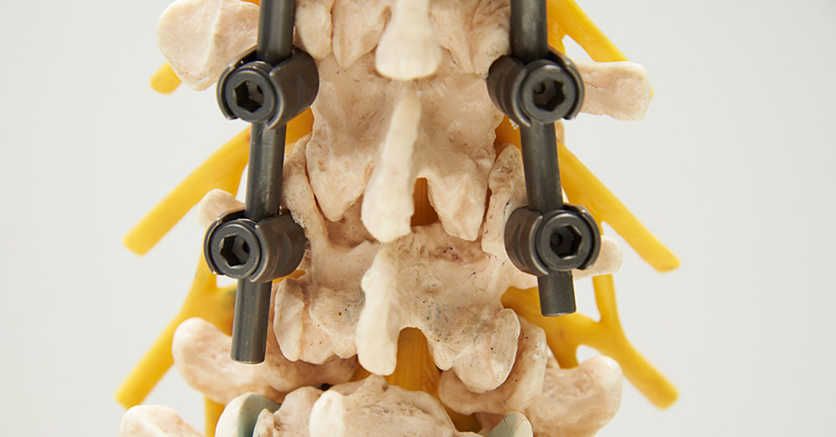
Percutaneous Pedicle Screw Fixation
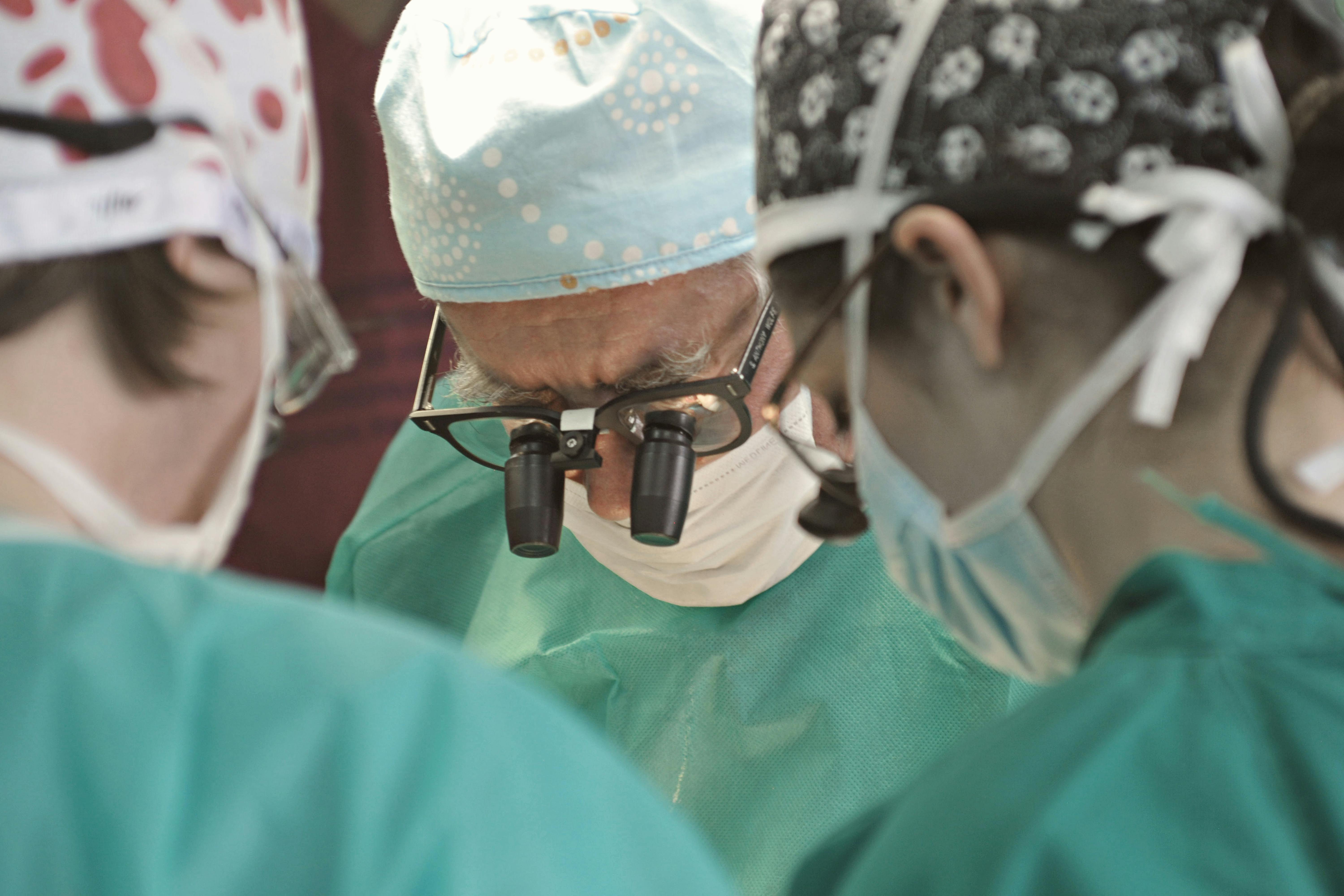
Percutaneous spinal stabilization is a revolutionary, minimally invasive spine surgery performed by Dr Shafeek in Calicut, designed to treat spinal instability from fractures, degenerative conditions, or tumors. This advanced technique uses specialized instruments and real-time imaging to place stabilizing screws and rods through small keyhole incisions. By avoiding large incisions and extensive muscle dissection, this approach reduces blood loss, minimizes post-operative pain, and shortens hospital stays. Patients benefit from immediate structural support and a faster recovery, making it one of the best treatments for debilitating spinal conditions.
Under general anesthesia, the patient is positioned face-down and multiple tiny incisions are made instead of a single large cut. Specialized instruments gently separate the back muscles, while real-time imaging such as fluoroscopy or computer navigation provides a detailed 3D view to guide screw placement with millimeter precision. Hollow pedicle screws are inserted over thin guidewires, and a stabilizing rod is tunneled beneath the skin to secure them, forming an immediate internal brace. The incisions are closed with minimal sutures, leaving only small scars. This technique restores spinal stability with far less trauma than open surgery, minimizes tissue damage and blood loss, and accelerates recovery. Navigated placement ensures strong, lasting fixation, offering excellent outcomes for spinal fractures and instability while enabling patients in Calicut to return to daily life sooner.
Recovery after percutaneous spinal stabilization in Calicut is significantly faster than traditional open fusion. Patients often experience less pain and are encouraged to walk within hours. Hospital stays are usually just a few days. A brace may sometimes be recommended briefly, while structured physical therapy helps regain core strength, flexibility, and function. This cutting-edge procedure offers excellent long-term spinal stability and pain relief, making it one of the best treatments in minimally invasive spine surgery under Dr Shafeek.